Pleural Mesothelioma Ct Scan - 29 28 Axial Ct Scan Of A Patient With A Right Sided Mesothelioma Download Scientific Diagram / Most people can go home as soon as the test is over.
Pleural Mesothelioma Ct Scan - 29 28 Axial Ct Scan Of A Patient With A Right Sided Mesothelioma Download Scientific Diagram / Most people can go home as soon as the test is over.. Increasingly, fdg pet/ct is used for diagnosis and management of malignant pleural mesothelioma. Diagnosis of malignant pleural mesothelioma is difficult because its early symptoms are commonly persistent cough, trouble catching your. Department of energy's office of scientific and technical information The extent of the pleural mass and particularly its spread into pleural fissures and along the mediastinal pleura are well seen with ct studies. Early detection of the fatal and incurable mesothelioma and the subsequent provision of radiation, surgical and palliative asbestosis treatments are known to help a patient to have the best possible chance to extend and improve the quality of life remaining.
ct scans are usually used for pleural mesothelioma and peritoneal mesothelioma, but not for the pericardial type of the disease. Ability to move air into and out of the lungs. These conditions include benign pleural diseases as well as metastasis of other tumors such as lung adenocarcinoma or chest wall sarcoma. To assess whether ct scans are a reliable tool for diagnosing pleural malignancy, researchers reviewed ct scans that had been routinely acquired for 345 patients included in the diaphragm study (isrctn10079972), a study of mesothelioma biomarkers that recruited patients suspected of having a pleural malignancy, from january 2014 to april 2016. If fluid is present in the chest cavity, the patient will undergo a pleural biopsy, where fluid or tissue is extracted and tested by a pathologist.
Modified Recist Criteria For Assessment Of Response In Malignant Pleural Mesothelioma Annals Of Oncology from els-jbs-prod-cdn.jbs.elsevierhealth.com To assess whether ct scans are a reliable tool for diagnosing pleural malignancy, researchers reviewed ct scans that had been routinely acquired for 345 patients included in the diaphragm study (isrctn10079972), a study of mesothelioma biomarkers that recruited patients suspected of having a pleural malignancy, from january 2014 to april 2016. Computerized tomography scan (ct scan) helps determine the location, size and extent of mesothelioma tumors and can help determine whether the tumor has invaded any of the adjacent structures. Imaging is important in diagnosing mesothelioma.it will provide information such as the extent of disease in the original organ and also show if the cancer has spread to. It is the main procedure used to diagnose pleural mesothelioma, and can be taken in two ways: Your general practitioner (gp) will assess your symptoms. Lim jh, choi jy, im y, et al.: Using a ct scan to diagnose mesothelioma. A biopsy is the only definitive way to confirm a mesothelioma diagnosis.
In one retrospective study, researchers reported 88% of malignant pleural mesothelioma patients showed pleural thickening on ct scan images.
The extent of the pleural mass and particularly its spread into pleural fissures and along the mediastinal pleura are well seen with ct studies. ct scan for pleural mesothelioma. pleural effusions are the first sign of pleural mesothelioma. Pulmonary function tests measure the lungs' These conditions include benign pleural diseases as well as metastasis of other tumors such as lung adenocarcinoma or chest wall sarcoma. See full list on radiopaedia.org. Suggest mesothelioma include an abnormal thickening of the pleura, calcium deposits on the pleura, fluid in the space between the lungs and the chest wall, or changes in the lungs themselves as a result of asbestos exposure. The ct scan is the primary imaging modality used to evaluate indicators of mesothelioma cancers. Pet/ct scan challenge of pleural effusion treatment for mesothelioma patients. Ultrasound image of the in vivo ncle procedure with the needle tip (n) and the confocal miniprobe (c) in a thickened pleura (p) of 1.5 cm. Your gp will conduct a physical examination and order tests. Computerized tomography scan (ct scan) helps determine the location, size and extent of mesothelioma tumors and can help determine whether the tumor has invaded any of the adjacent structures. pleural mesothelioma ct scan :
ct scan for mesothelioma a ct scan generates detailed three dimensional images of specific regions of the body. See full list on radiopaedia.org. The workers developed the condition anywhere from 3 to 34 years after asbestos exposure. Doctors can typically find masses on the lungs or pleura, but these diagnostic tests cannot always determine whether the mass represents cancer or a benign pleural plaque. The main test to stage mesothelioma is a ct scan.
1 from Biopsies if imaging scans reveal signs of pleural mesothelioma, a pathologist will perform a biopsy, which is a procedure that removes a small fluid or tissue sample from a patient's body to test it for. Findings that could indicate mesothelioma is a thickening of the pleura, deposits of calcium on the pleura, fluid in the space between the chest wall and lungs, and changes in the lungs themselves from exposure to asbestos. A biopsy is needed to confirm a diagnosis of pleural mesothelioma. The main symptoms are shortness of breath, pain when breathing, chest/shoulder/upper arm pain, loss of appetite, weight loss, and persistent cough or bouts of pneumonia. The journal of thoracic disease indicates that not only does mesothelioma show up on a ct scan but it is the preferred diagnostic tool of choice for advanced stage mesothelioma cases. The aim of this study is to propose a diagnostic aid system that is capable of segmenting and measuring the pleural thickening caused by a pleural disease called "malignant pleural mesothelioma". The workers developed the condition anywhere from 3 to 34 years after asbestos exposure. ct scan for mesothelioma a ct scan generates detailed three dimensional images of specific regions of the body.
Helps to predict lung function/capacity after surgery.
Pet and mri scans provide even further detail and may be used to differentiate pleural thickening from malignant mesothelioma. $30 billion dollars is available.jan 01, 2004 · ct is the primary imaging modality used for the evaluation of mpm. If the cancer is in the chest, the doctor may perform a thoracoscopy. The association between pleural plaques and pleural mesothelioma remains controversial. Findings that could indicate mesothelioma is a thickening of the pleura, deposits of calcium on the pleura, fluid in the space between the chest wall and lungs, and changes in the lungs themselves from exposure to asbestos. ct scans are usually used for pleural mesothelioma and peritoneal mesothelioma, but not for the pericardial type of the disease. pleural effusions are the first sign of pleural mesothelioma. Doctors also use mesothelioma blood tests to measure treatment response. The journal of thoracic disease indicates that not only does mesothelioma show up on a ct scan but it is the preferred diagnostic tool of choice for advanced stage mesothelioma cases. The workers developed the condition anywhere from 3 to 34 years after asbestos exposure. ct is particularly valuable in assessing the extent of malignant mesothelioma as well as establishing the presence of pleural effusion or parenchymal disease obscured by a pleural effusion. Helps to predict lung function/capacity after surgery. Scar tissue forms as the result of asbestos fibers puncturing the mesothelial cell lining.
Using a ct scan to diagnose mesothelioma. Doctors can typically find masses on the lungs or pleura, but these diagnostic tests cannot always determine whether the mass represents cancer or a benign pleural plaque. Your gp will conduct a physical examination and order tests. Helps to predict lung function/capacity after surgery. If the cancer is in the chest, the doctor may perform a thoracoscopy.
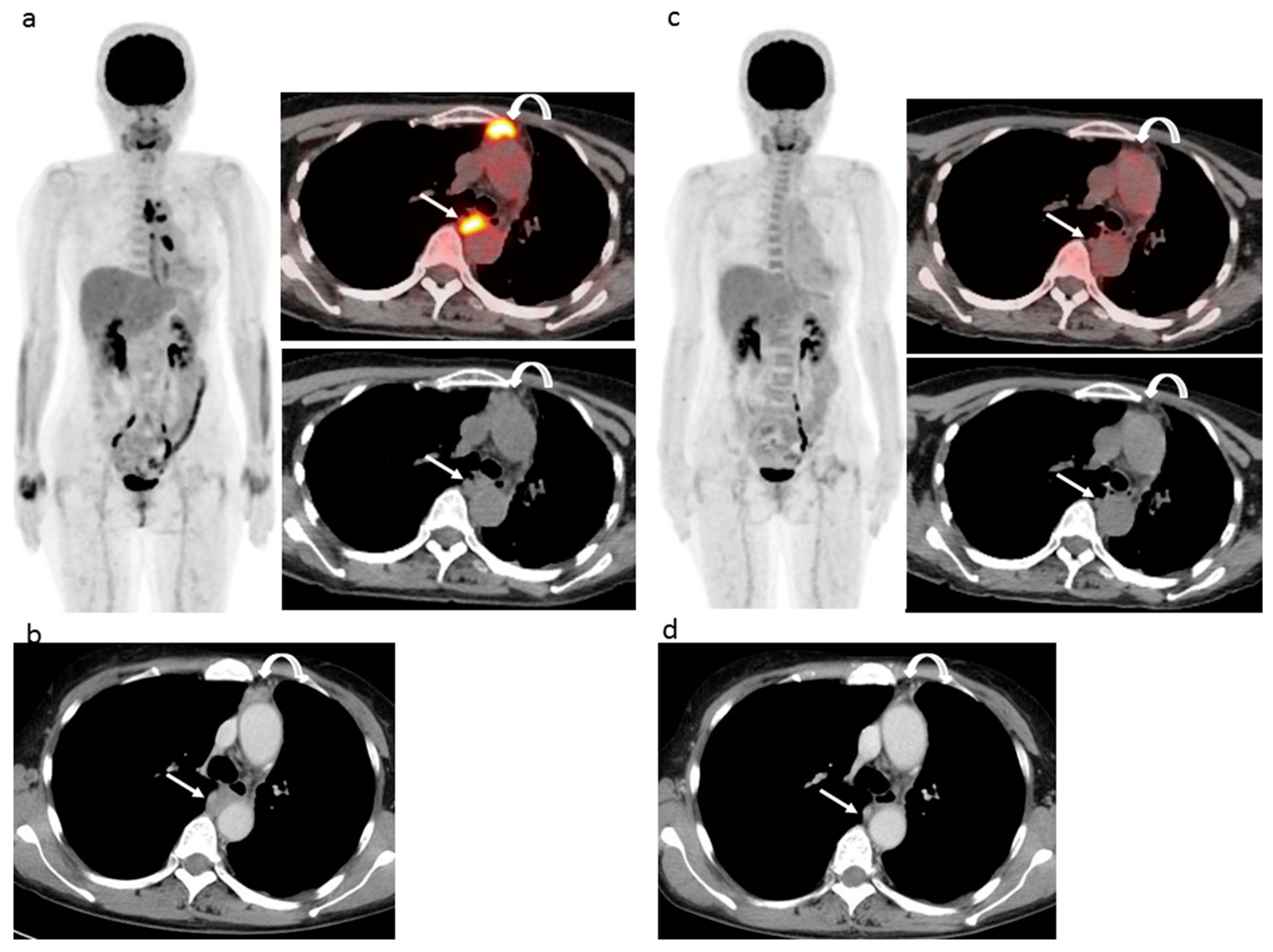
Cancers Free Full Text Response To Immune Checkpoint Inhibitor Therapy In Patients With Unresectable Recurrent Malignant Pleural Mesothelioma Shown By Fdg Pet And Ct Html from www.mdpi.com Scar tissue forms as the result of asbestos fibers puncturing the mesothelial cell lining. Role of ct scans suggest or rule out a mesothelioma diagnosis the journal of thoracic disease indicates that not only does mesothelioma show up on a ct scan, but it is the preferred diagnostic tool of choice for advanced stage mesothelioma cases. ct scan for mesothelioma a ct scan generates detailed three dimensional images of specific regions of the body. Department of energy's office of scientific and technical information The only known cause of pleural mesothelioma is genetic damage caused by exposure to asbestos fibers. A biopsy is needed to confirm a diagnosis of pleural mesothelioma. Pet/ct scan challenge of pleural effusion treatment for mesothelioma patients. Computed tomography (ct) scan in a male veterans administration patient with a history of asbestos exposure and an enlarging abdominal girth.
Findings that could indicate mesothelioma is a thickening of the pleura, deposits of calcium on the pleura, fluid in the space between the chest wall and lungs, and changes in the lungs themselves from exposure to asbestos.
Multiple intracranial metastases from postoperative giant sarcomatoid malignant pleural mesothelioma a case report and literature review : A biopsy is needed to confirm a diagnosis of pleural mesothelioma. A ct scan or an mri may also be useful. The ct scan is the primary imaging modality used to evaluate indicators of mesothelioma cancers. This is due to the details provided in the images of the peritoneum (the lining of the abdomen) and the pleura (the lining of the lung). Most people can go home as soon as the test is over. pleural thickening is the thickening of the lung lining, or pleura. pleural effusion occurred in 74% of those patients. The workers developed the condition anywhere from 3 to 34 years after asbestos exposure. Diagnosis of malignant pleural mesothelioma is difficult because its early symptoms are commonly persistent cough, trouble catching your. ct scan for pleural mesothelioma. Early detection of the fatal and incurable mesothelioma and the subsequent provision of radiation, surgical and palliative asbestosis treatments are known to help a patient to have the best possible chance to extend and improve the quality of life remaining. Malignant pleural mesothelioma needs to be distinguished from other conditions.
Early detection of the fatal and incurable mesothelioma and the subsequent provision of radiation, surgical and palliative asbestosis treatments are known to help a patient to have the best possible chance to extend and improve the quality of life remaining mesothelioma ct scan. This is due to the details provided in the images of the peritoneum (the lining of the abdomen) and the pleura (the lining of the lung).


0 Comments